A 2d echo with a Doppler is also known as a two-dimensional Echocardiography with Doppler. It is a standard medical scanning test. Echocardiography with Doppler uses ultrasound technology to give your doctor a live-moving picture of your heart. It does not carry any radiation, unlike x-ray and CT scans. Hence it can be done even to pregnant women. It is a noninvasive test and also a pain-free test. It takes less than a few minutes. 2d echo test with Doppler helps to detect a variety of heart-related disorders. 2d echo with Doppler is available at many hospitals in Hyderabad, such as Apollo hospital, Care hospital, Yashoda hospital, and TX hospital.
What are the Uses of a 2d Echo with Doppler?
The application of 2d echocardiogram with Doppler in clinical practice is broad, and a few of them includes:
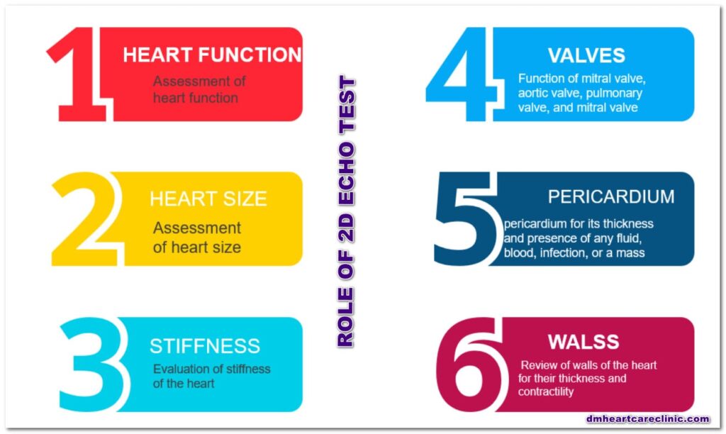
- Assessment of heart function
- Assessment of heart size
- Review of the location of the heart
- Study of dimensions of four chambers of the heart
- Assessment of sizes of various vessels in the vicinity of the heart like aorta, pulmonary artery, and superior vena cava. Inferior vena cava and pulmonary veins
- Evaluation of the function of four valves of the heart a mitral valve, aortic valve, pulmonary valve, and mitral valve
- Assessment of pericardium for its thickness and presence of any fluid, blood, infection, or a mass
- Review of walls of the heart for their thickness and contractility
- Assessment of septum of the heart
- Evaluation of stiffness of the heart
APPLICATION
2d echo test or heart scan is helpful for the evaluation of anatomy, whereas doppler helps assess the speed of the blood and function of four valves of the heart.
Various diseases where there is a role of transthoracic echocardiography (TTE) with Doppler is
Coronary artery disease or CAD
Arrhythmias or heart rhythm abnormalities
Cardiomyopathy
- Hypertrophic cardiomyopathy or HCM
- Restrictive cardiomyopathy or RCM
- Dilated cardiomyopathy or DCM
Heart failure or Congestive heart failure
Problems with the valves or faulty heart valves
- Mitral stenosis or MS
- Mitral regurgitation or MR
- Aortic stenosis or AS
- Aortic regurgitation or AR
- Pulmonary stenosis or PS
- Pulmonary regurgitation or PR
- Tricuspid stenosis or TS
- Tricuspid regurgitation or TR
Disorders of pericardium
- Pericardial effusion
- Constrictive pericarditis
Tumors of the heart
A clot in the heart
Infections in the heart
- Infective endocarditis
- Myocarditis
- Pericarditis
Congenital heart disease
- Atrial septal defects or ASD
- Ventricular septal defects or VSD
- Atrioventricular septal defects or AVSD
- Patent ductus arteriosus or PDA
- Tetralogy of Fallot or TOF
- Coarctation of the aorta
Hypertensive heart disease
Pulmonary artery hypertension (PAH)
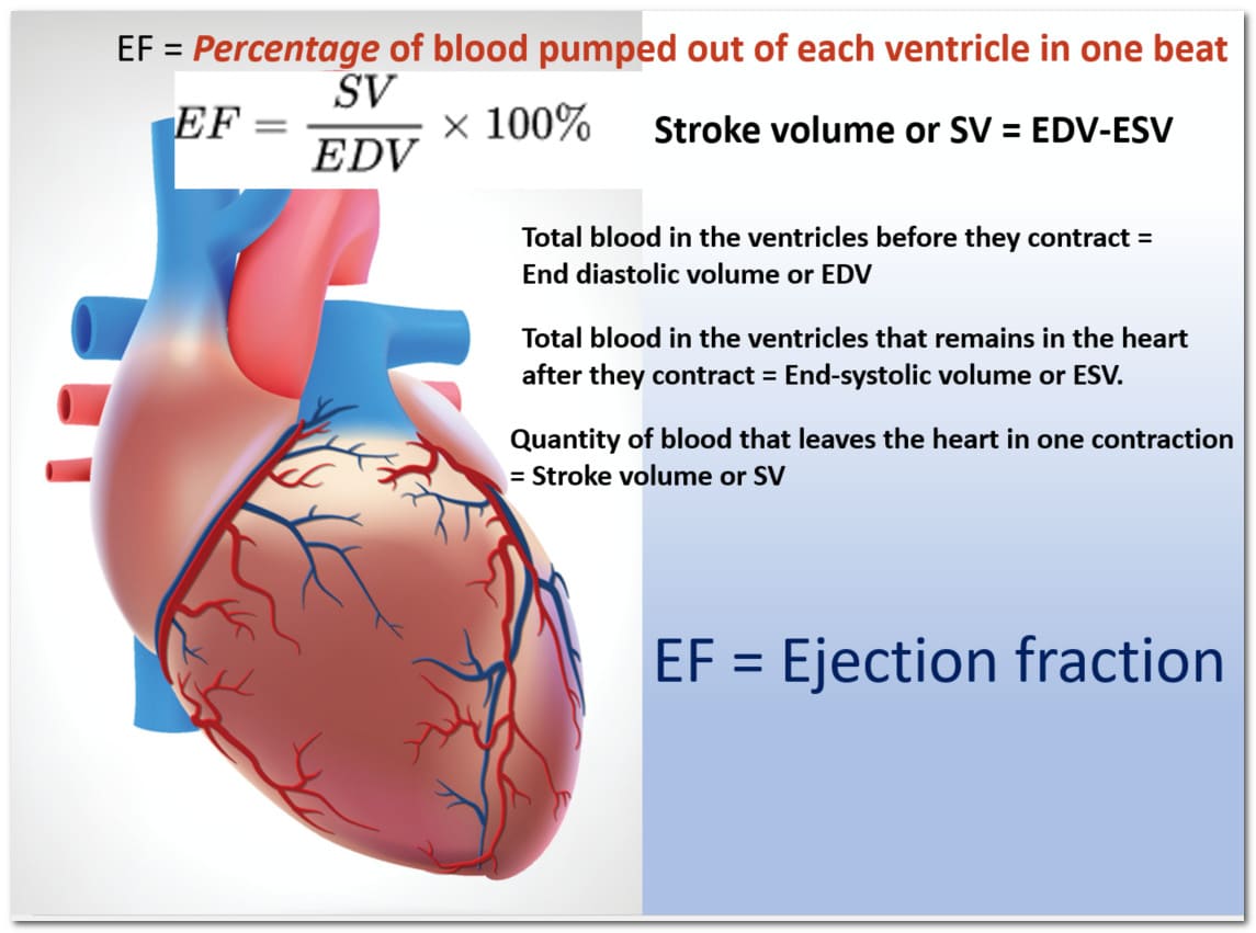
Assessment of cardiac output or ejection fraction
Assessment of the fluid status of the body
Indications
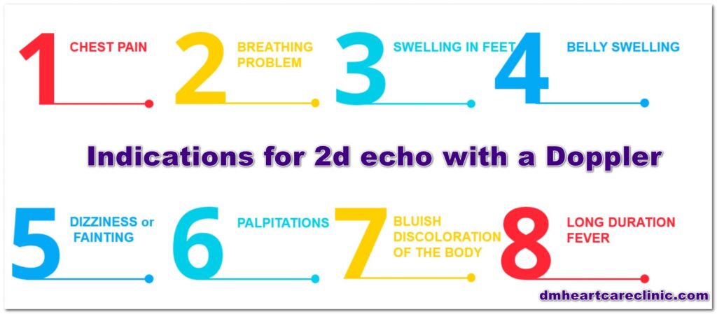
An echocardiogram with Doppler is indicated if you have the following complaints.
- Chest pain
- Breathlessness /dyspnea
- Swelling in feet
- Swelling of the abdomen or stomach swelling
- Dizziness
- Syncope or fainting
- Heart Palpitation
- Bluish discoloration of the body or cyanosis
- Long duration of fever
How Much Does a 2D Echo with a Doppler Cost?
The cost of a 2d echocardiogram with Doppler in a DM heart care clinic is Rs 1000.
How is a 2d echo test done?
Before lying down on a bed for a 2d echo test, you’ll be asked to take off any clothes that cover your upper half. During the test, you may be given a hospital gown to wear. Several small sticky sensors, known as electrodes, will be applied to your chest while you are lying down. These will be attached to a machine that will track your heartbeat during the exam.
Your chest will be lubricated with a lubricant gel, or the ultrasonic probe will be lubricated directly. The probe will be moved across your chest as you lie on your left side. A connection connects the probe to a nearby machine that will display and record the images it generates.
The sound waves created by the 2d echo probe will not be audible, although you may hear a swishing noise during the 2d echo scan. This is normal and is simply the sound of the probe picking up the blood flow via your heart. The 2d echo test will usually take 15 to 60 minutes, and you will be able to return home soon after.
2D Echo test at home?
A two-dimensional echocardiogram needs an ultrasound machine to perform. In India, it is not legal to move an ultrasound machine from the place of registration. Hence 2d echo test is not done at home in India.
2D echo test normal report
To say what is normal in 2d echo takes more than 100 pages to write. In general, a 2d echo report is considered normal if
- The size of the heart is normal
- The function of the heart is normal
- The ejection fraction is normal
- The thickness of the walls of the heart is normal
- The size of all four chambers of the heart is normal
- The size of the aorta and pulmonary artery is normal
- Size of inferior vena cava and superior vena cava is nirmal
- All four valves of the heart are opening and closing properly
- There is no pulmonary arterial hypertension
- Stiffness of the heart is acceptable
- No mass or vegetation in the heart
- No clot in the heart
- No fluid in the sac surrounding the heart
- There is no RWMA
- No hole in the heart
- All chambers are located in a correct location
- The heart is situated in a current location
If the 2d echo is normal, is my heart ok?
The 2d echo test is one of the many tests available for the heart. It is an imaging test for the heart and checks for the anatomy and function of the heart. You can have multiple blockages in your coronary arteries with a normal 2d echo test. So it is always preferable to combine ECG, 2d echo test, treadmill test, and medical history to make a diagnosis.
ECG vs. 2d echo: Which is better
Both the tests are very different from each other. They are complementary to one another but not competitors to each other. Both are better in their ways.
For heart rate, rhythm abnormalities, and ischemia- ECG is better
Heart function, size, congenital heart diseases, valve problems- 2d echo is better
TMT vs. 2d echo: Which is better
Again, there is no comparison between the two. The applications and uses of the two tests are entirely different. Your doctor decides the proper test for you.
Heart function, size, congenital heart diseases, valve problems- the 2d echo is better.
To diagnose blockages in the heart- the TMT test is better
What can an echocardiogram miss?
An echocardiogram can miss certain pathologies.
- Abnormalities of coronary arteries such as (require angiogram)
-
-
-
-
-
- Blockages
- Dilatations
- Unusual location
- Unusual number
-
-
-
-
-
- Posterior pathologies (may require TEE test)
- Small vegetation and clots (may require contrast 2d echo studies)
- May miss small heart attacks
- May miss chronic stable angina (stress 2d echo is required)
- May miss clots in the lungs or pulmonary embolism (requires CT scan)
What are the types of 2d echo tests?
There are several types of 2d echo tests in cardiology
- 2d echo test with a Doppler
- TEE test or transesophageal echocardiogram
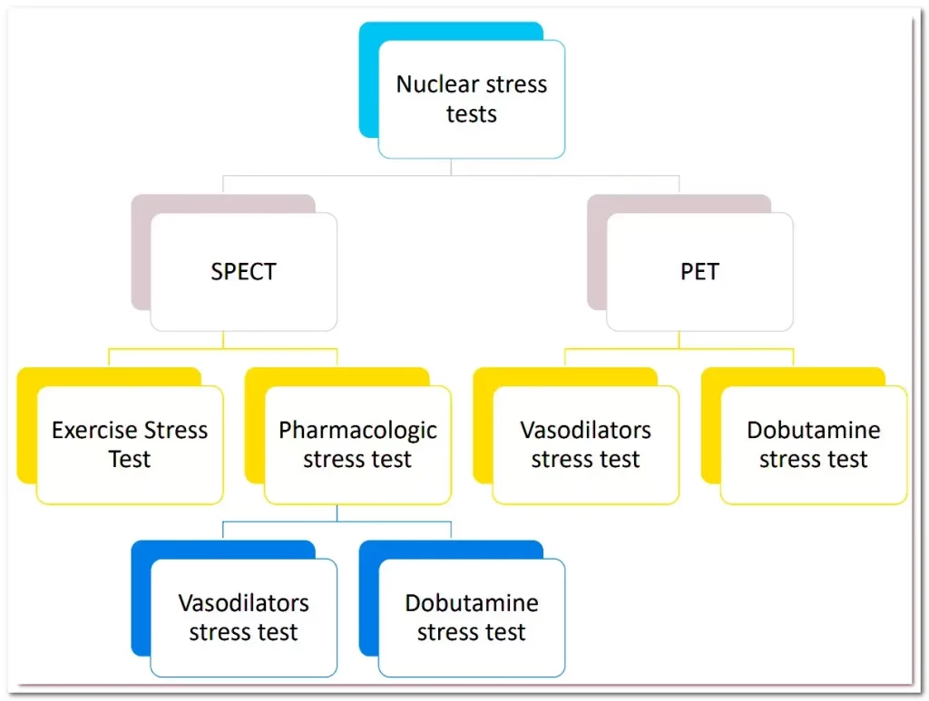
- Stress echo test
- Fetal 2d echo test
- 2d echo test with a Doppler and strain imaging
- Contrast 2d echo
Stress 2d echo test
You’ll exercise on a treadmill or stationary bike while your doctor measures your blood pressure and heart rhythm during a stress echocardiogram.
Your doctor will capture ultrasound images of your heart when your heart rate hits its peak levels to see if your heart muscles are getting enough blood and oxygen as you exercise.
If you have chest pain that your doctor suspects are caused by coronary artery disease or a heart attack, they may prescribe a stress echocardiography test. In cardiac rehabilitation, this test will also evaluate how much activity you may safely endure.
Contrast 2d echocardiography
Contrast echocardiography (commonly known as “a contrast echo”) is an imaging technique that takes photos of the heart while it is beating using sound waves (ultrasound). A special dye (contrast agent) is injected into your vein during the test to help display structures in your heart more clearly. It permits ultrasound images of the inside of the heart to be seen more clearly. A contrast echo is most commonly used to look for heart issues that standard 2d echo procedures can’t detect. The test takes about 60 minutes from start to finish.
Fetal 2d echocardiogram
A fetal echocardiogram is a type of ultrasound used to examine the heart of a fetus. It’s usually done between weeks 18 and 24 of the second trimester. Your doctor will be able to view the anatomy and function of your unborn child’s heart with this test.
Transesophageal 2d echocardiogram
A transesophageal echocardiogram test is done using an echo probe passed via the mouth and throat into the esophagus. The transesophageal echocardiogram test takes a continuous picture of the heart while it beats, which is then transferred to a monitor that is close by. The esophagus is the food pipe connecting the mouth with the stomach esophagus. The transesophageal echocardiogram probe takes the images of the heart from the back of the heart.
2d echo test vs. 3d echo test?
Most patients need a 2d echo test for their heart diseases. Usage of 3d echo is minimal in cardiology. Only a subset of patients requires a 3d echo test. 3 d echo test is expensive but not necessarily superior to a 2d echo test. The 3d echo test requires more expertise. 3d echo is mainly used for research purposes, and some exceptional cases like atrial septal defects, and accurate measurement of heart function
2d echo test in pregnancy
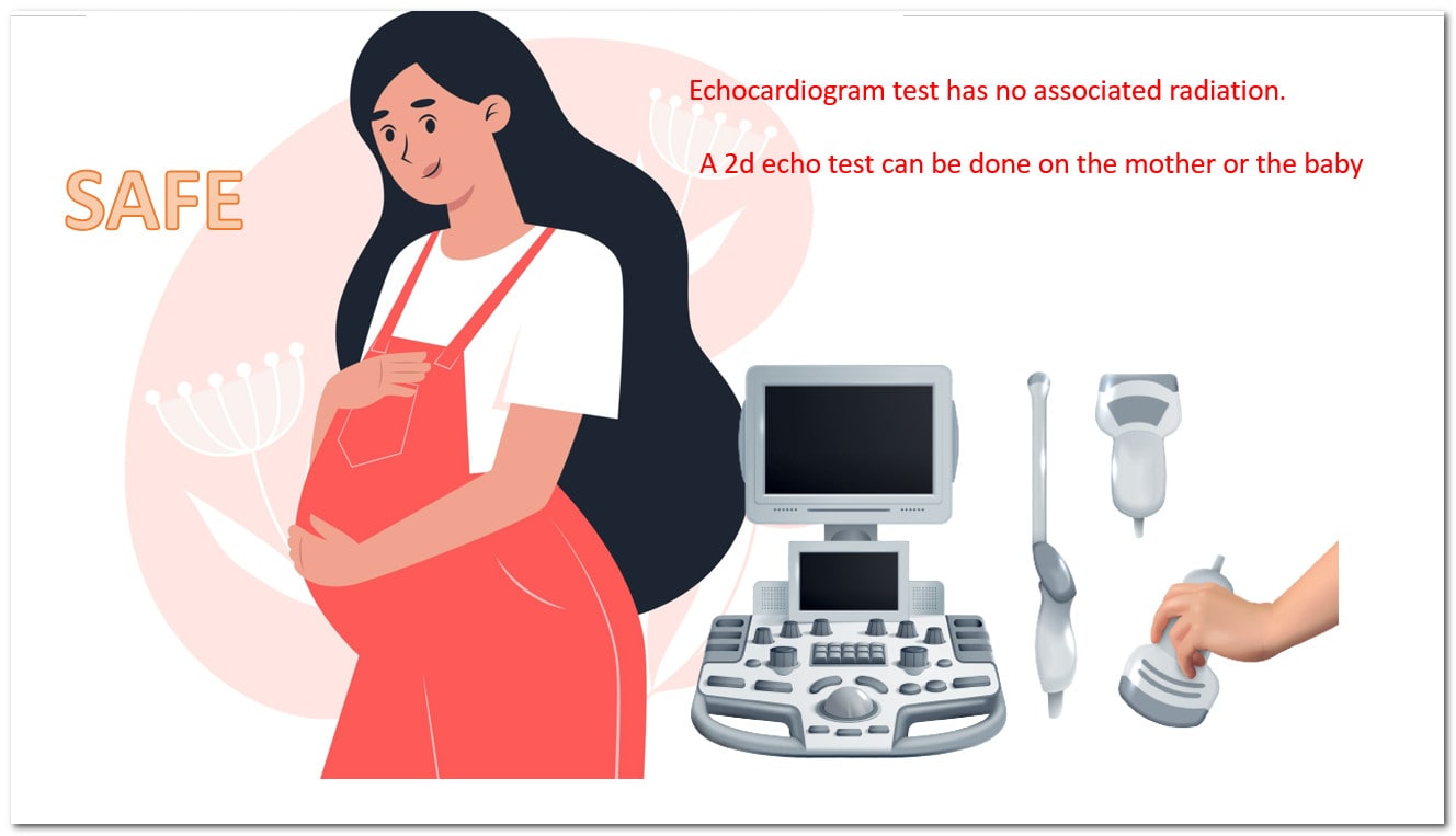
2d echo test can be done during pregnancy also. It does not cause any radiation to the baby. During pregnancy, a 2d echo can be done on the mother or baby or on both.
Conclusion: Why You Should Get Your Next 2d Echo Test
A two-dimensional echocardiogram is a non-invasive diagnostic test used to measure the heart’s pumping function. It is also known as a 2d echocardiogram. It is available in many hospitals in Hyderabad. Why wait for a heart attack? Get your heart tested today!
DM HEART CARE CLINIC, Attapur, Hyderabad, offers 2d echo tests in Hyderabad at an affordable price. The cost of a 2d echo test starts from 600 rupees.
Click here for a 2d echo test in Telugu






Pingback: Atrial septal defect - ECG 2D ECHO TMT CARDIOLOGIST HYDERABAD
Pingback: HEART ATTACK IN YOUNG PEOPLE: RISK FACTORS, TESTS, AND TREATMENT - ECG 2D ECHO TMT CARDIOLOGIST HYDERABAD
Pingback: The Complete Guide to Myocardial Infarctions (aka Heart Attacks) - CARDIOLOGIST HYDERABAD
Pingback: Left hand pain – 10 Reasons you should know - ECG 2D ECHO TMT CARDIOLOGIST HYDERABAD
Pingback: Ejection fraction of the heart: It is not just a number - ECG 2D ECHO TMT CARDIOLOGIST HYDERABAD
Pingback: HEART HEALTH CHECK-UP PACKAGES IN HYDERABAD - ECG 2D ECHO TMT CARDIOLOGIST HYDERABAD
Pingback: ELECTROCARDIOGRAM TEST IN TELANGANA - ECG 2D ECHO TMT CARDIOLOGIST HYDERABAD
Pingback: Cardiologist in Mehdipatnam Hyderabad - DM HEART CARE CLINIC
Pingback: 2d echo test in newborn baby - DM HEART CARE CLINIC
Pingback: 2d echo test in pregnancy
Pingback: 2d echo test in Attapur - DM HEART CARE CLINIC