An electrocardiogram is a common cardiac test to study the heart’s electrical activity.
Introduction to an electrocardiogram or electrocardiography
An electrocardiogram is a standard test that most doctors can administer at the office or outpatient center without requiring pricey equipment. Because of this, it’s one of the most common tests used by medical professionals to keep track of the overall health of their patients. An electrocardiogram, or EKG, monitors electrical activity in your heart.
An electrocardiogram is a necessary medical test used to monitor your heart health. It’s also known as an EKG or ECG. Either a technician or a cardiologist (heart specialist) conduct an ECG test. ECG can study the speed at which your heart is beating. Electrodes are attached to your chest and arms during the procedure, allowing doctors to gather information about your heart’s electrical activity.
If you consider an electrocardiogram test in Telangana, you may have some questions about how it works and what happens before, during, and after the ECG test. This article will cover everything you need to know about the electrocardiogram test and what happens during one.
How does electrocardiography or EKG work?
An electrocardiogram, or EKG, monitors electrical activity in your heart. During a routine checkup with your doctor, you can expect to undergo an EKG test. You’ll lie on a table while electrodes are placed on your chest to measure heartbeats.
Types of electrocardiogram or ECG?
Based on the number of tracings we get, ECG is classified as below.
12 lead electrocardiogram
It is by far the most common ECG type done in medical practices. It takes the information from 12 different angles and gives it as 12 tracings. 10 leads to attached to your body (4 to the four limbs and 6 to your chest). It produces more accurate and reliable results.
Six lead ECG
It takes the information from 6 angles and gives it as six tracings.
Two lead ECG
It takes the information from 3 angles and gives it as two tracings.
One-Lead electrocardiogram
For a single channel, one Lead ECG, one electrode is attached to the Left Leg (LL) and another to Right Arm (RA). Only one tracing is what we get.
Depending on the duration of the study. ECG is classified as
1. Resting electrocardiogram or ECG
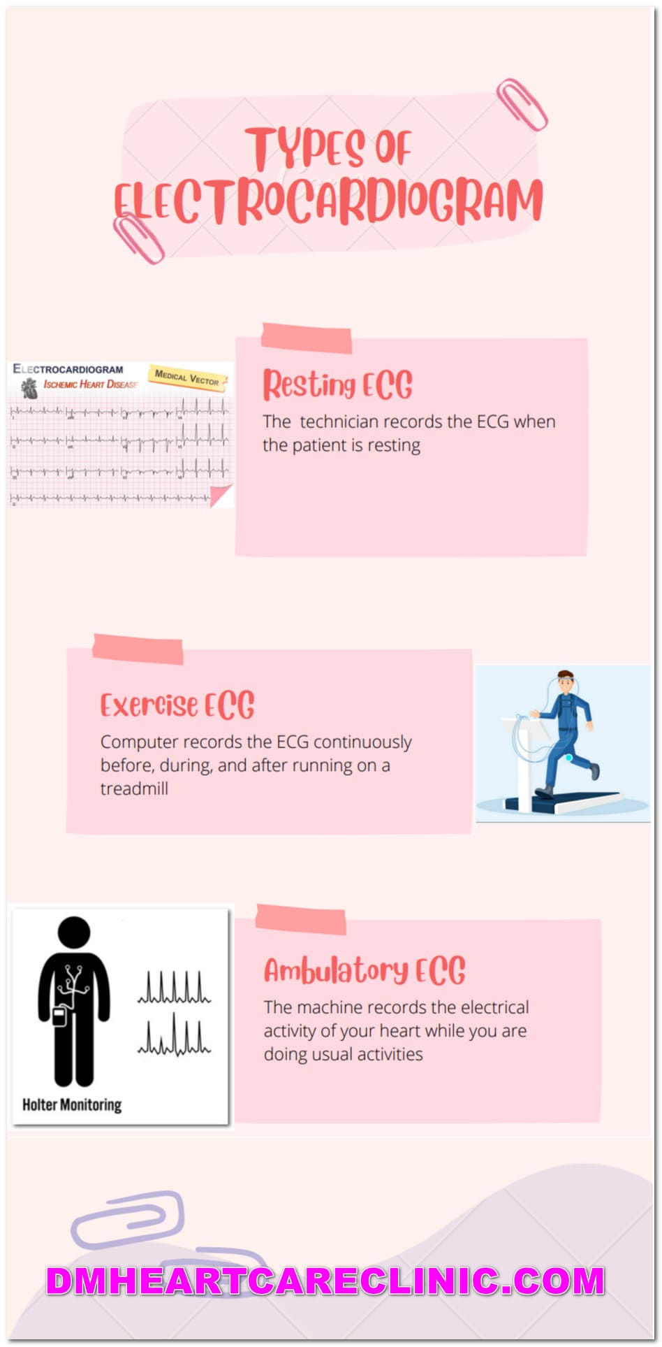
Resting ECG is nothing but a routine ECG. It records an ECG of the heart for a minute or 2 minutes. The doctor or technician records the ECG when the patient is resting.
2. Exercise ECG or stress ECG
The exercise ECG test is a non-invasive and inexpensive medical test to diagnose coronary artery diseases. It is a cardiac test where a computer records the ECG continuously before, during, and after running on a treadmill. ECG is recorded for 5 to 20 minutes. Coronary artery disease happens when the arteries that supply the heart get narrowed.
3. Ambulatory ECG
When the machine records the electrical activity of your heart while you are doing usual activities, it is referred to as Ambulatory ECG. It can be
A. Holter recording
A Holter monitor is a tiny, wearable medical device that records the heartbeat. It determines if you’re having heart rhythm abnormalities(arrhythmias). A typical Holter test records ECG for 24 hours to 72 hours.
Suppose a resting ECG does not provide adequate information for palpitations or fainting. In that case, your cardiologist may advise a Holter.
B. Event monitor
This device only records your ECG when symptoms appear, not before. You switch on the device and insert the electrodes on your chest to capture the ECG when you experience symptoms. This device is carried in your pocket or worn on your wrist.
If you have heart palpitation and or fainting, which occurs once a month or so, your doctor prescribes an Event monitor.
C. Loop recorder
A loop recorder is a tiny chest monitor implanted beneath the chest’s skin. It can be left in place for three years or longer to monitor cardiac rhythms. Your doctor advises this test when Holter’s report is inconclusive, and you have persistent symptoms that occur pretty infrequently.
.
Why would you get an EKG or ECG?
An electrocardiogram, or EKG, records electrical activity in your heart. These signals can be affected by many medical conditions, so your doctor may order one as part of a routine checkup or to rule out certain conditions.
Symptoms of heart disease are:
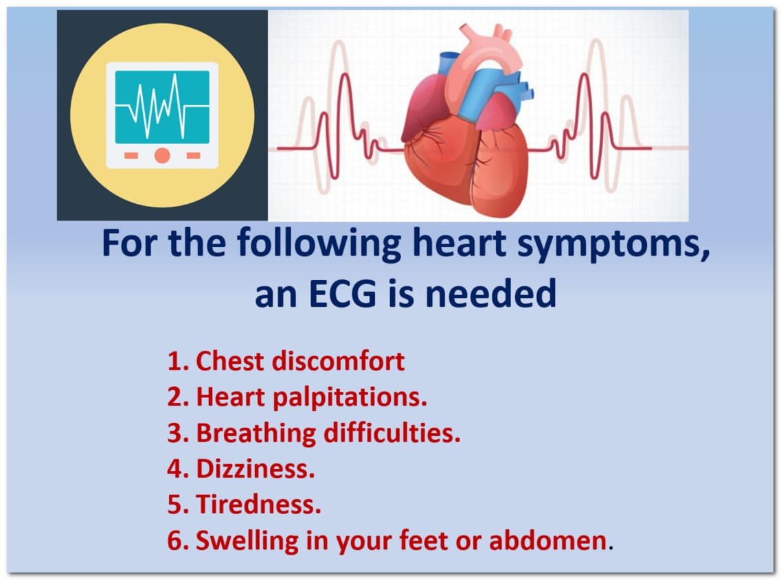
If you are having the following heart symptoms, your cardiologist may recommend an ECG.:
- Chest discomfort or left arm pain.
- Heart palpitations.
- Breathing difficulties.
- Dizziness.
- Tiredness.
- Swelling in your feet or abdomen.
A cardiologist may recommend an ECG for those who are at risk of heart disease, such as:
- Elevated blood pressure or hypertension.
- Increased blood cholesterol levels or hyperlipidemia.
- Diabetes mellitus.
- Smoking.
- Age -As you become older, you’re more likely to have damaged and restricted arteries and a weakened or thicker heart muscle.
- Male Sex. Male are at higher risk of heart disease than females.
- Family history of premature heart diseases.
- Unhealthy diet.
- Obesity.
ECG is also done as a routine health checkup or pre-operative evaluation for non-cardiac surgery.
How long does ECG or electrocardiogram take?
Typically, electrocardiograms (EKGs) can be performed in 3 to 5 minutes. But don’t worry—some people are done with their EKG within 2 minutes! People who have pacemakers or other electrical devices may need more time for their EKG readings to be evaluated by a cardiologist.
How is an ECG or electrocardiogram performed?
The electrocardiogram, or EKG, has been a fundamental part of cardiovascular care for more than 100 years. An EKG measures your heart’s electrical activity as it pumps blood through your arteries. Electrodes stuck to your skin pick up the heart’s electrical activity and give it as tracings on graph paper. An EKG may also help detect any heart conditions leading to sudden cardiac arrest and death.
- You will be informed about the ECG procedure.
- You may be changed to a hospital gown before the ECG test.
- You asked to lie down on a bed or a table.
- A sticky gel was applied to specific areas over all the limbs and chest areas as specified by the doctor.
- Your technician or a heart specialist connects 12 electrodes ( knobs or patches) over the place where the gel is applied. The wires from the electrodes have connected to a device called an ECG machine. ECG machine will start recording the electrical activity and gives the result in graphical form. The report from the ECG machine can be printed or shared with you wirelessly.
- You can breathe during the exam, but you must remain completely still. Make sure you’re warm and comfortable before lying down. Moving, talking, or shivering may cause the test to fail.
Are there any risks with an electrocardiogram test?
No, there are very few risks with an electrocardiogram. It’s considered a completely safe procedure and has no side effects.
What is the information the doctor gets from an ECG test?

1. Heart rate
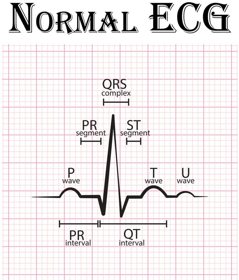
ECG test can accurately compute Heart rate. It can determine if your heartbeat is regular or irregular. The heart got four chambers- two upper chambers and two lower chambers. Upper chambers (atria) produce one spike (p wave), whereas lower chambers( ventricles) produce a separate spike (called QRS wave).
Each p wave (generated by the upper chambers) is usually followed by a QRS wave (produced by upper chambers). This pattern is called normal sinus rhythm. The number of p waves in one minute is the rate of upper chambers in one minute. Similarly, the number of QRS waves in one minute is the rate of lower sections in one minute.
Normally
Number of p waves = Number of QRS waves
Rate of upper chambers = Rate of lower chambers
Sometimes, there may not be coordination between the heart’s upper chambers and lower chambers. The upper chamber can beat slower or faster than the lower chambers of the heart.
ECG can individually study the rate of upper and lower chambers, which is impossible with pulse oxymetry or physical examination.
Ecg can detect heart rate, rate of upper chambers, and rate of the lower chambers.
Slow heart rate: Heart rate less than 50 beats per minute
Fast heart rate: Heart rate of more than 100 beats per minute
Premature heartbeats: Extra heartbeats that arise from an abnormal location
2. Heart rhythm abnormalities
Sometimes there can be several p waves for one QRS or vice versa, both abnormal. We call them arrhythmias or heart rhythm abnormalities. An ECG test can quickly detect these anomalies, which is not achievable with a 2d echo or a heart angiography. Sometimes p waves are absent, which is also abnormal.
ECG can detect Rapid, slow, or irregular heartbeats. ECG helps diagnose heart rhythm abnormalities like
- Supraventricular tachycardia: P = QRS
- Atrial fibrillation (A-fib): No P waves
- Atrial flutter: Flutter waves than normal P waves
- atrial tachycardia: Abnormal P waves
- Ventricular tachycardia: QRS> P waves, Regular QRS
- Ventricular fibrillation: QRS> P waves, Irregular QRS
Slow heartbeat (bradycardia)
- Sick sinus syndrome: Slow P wave rate
- Conduction block:
3. Heart attack or myocardial infarction
- ECG will be the first test your heart doctor recommends if you get chest pain. When a part of the heart is deprived of blood supply due to a heart attack, it produces specific characteristic changes in the ECG. These are hallmarks of a heart attack and can be easily identified by a heart specialist.
- ECG can show an old heart attack or a heart attack currently happening.
- ECG is also helpful in assessing the severity of the heart attack, part of the heart affected, and duration of the heart attack.
- ECG can also estimate a patient’s prognosis with a heart attack.
- Even complications that may arise due to a heart attack such as very fast heart rates (ventricular tachycardia, ventricular fibrillation, or atrial fibrillation), slow heart rates (sinus bradycardia or complete heart block)
- ECG plays a more vital role than any cardiac test regarding heart attack.
- The ECG patterns may aid in determining which section of the heart has been affected and how severe the damage is.
4. Blood and oxygen supply to the heart.
- Not only a heart attack, but there are also other causes for the decreased supply of blood to the heart muscle. This is called ischemia. Ischemia can produce chest pain which comes and goes. If an ECG is performed when the patient is experiencing chest pain, ischemia changes can be seen.
5. Heart structure changes.
- An ECG can provide hints about
- enlargement of the heart
-
-
- enlargement of the upper chambers of the heart
- enlargement of the lower chamber of the heart
-
-
- Increased thickness of the heart muscle
- Congenital heart defects like holes in the heart such as
-
-
-
- Atrial septal defect or ASD
- Ventricular septal defect or VSD
- Patent ductus arteriosus or PDA
- Tetralogyogy of Fallot or TOF
- Ebstein anomaly
-
-
- Raised pressure in the heart chambers like pulmonary hypertension or systemic hypertension
- Faulty valves of the heart like mitral stenosis, aortic stenosis, aortic regurgitation, mitral regurgitation
- , abnormal position of the heart, like the right-side heart
6. Inflammation of the heart
- pericarditis and myocarditis can produce specific ECG changes
7. Congenital electrical disturbances of the heart
- Some genetic issues in the heart’s conducting system increase the risk of sudden death or arrhythmias. ECG can give clues for such diseases
-
-
-
-
- Long qt syndrome
- Short qt syndrome
- Brugada syndrome
- WPW syndrome
- Early repolarisation syndrome
-
-
-
-
8. Electrolyte disturbances in the body
Disturbances in the body’s electrolytes produce characteristic ECG changes. ECG can sometimes give the first clue for underlying abnormality. Low potassium, high potassium, low calcium, and high calcium produce their findings in the ECG. An experienced heart doctor can make the diagnosis of underlying Electrolyte disturbances.
A doctor will use a stethoscope, or even just their ears, to listen to your heart. An EKG can also catch abnormalities such as arrhythmias (abnormal heart rhythms) and make it easier for doctors to see if you have a heart attack.
Where will I get my EKG done in Hyderabad, Telangana?
Your doctor will either perform your EKG in their office or refer you to a cardiologist ( heart doctor) for further testing to get your EKG done.
Dr. Malleswara Rao is a reputed heart specialist for interpreting EKG or ECG. Consult our heart doctor in Hyderabad for your heart diseases.
Other cardiac tests for heart diseases if your ECG is abnormal or inconclusive
- Two-dimensional echocardiogram (2D Echo)
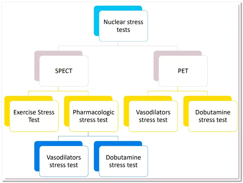
- Treadmill test
- CT calcium score
- CT coronary angiogram
- Lipid profile
- Serum homocysteine
- Lipoprotein A (Lp(a))
- High-sensitivity C-reactive protein or Hs CRP
- Troponin blood test
ECG or electrocardiogram test price in Hyderabad, Telangana?
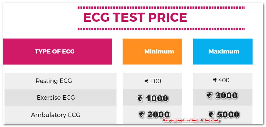
The cost of a resting ECG ECG test typically starts from 150 Indian rupees in Hyderabad. It can go up to 300 rupees in Hyderabad.
The cost of ambulatory ECG is expensive when compared to resting ECG. It will be around 2000 to 3000 for 24 hours and up to 5000 for 72 hours,
Exercise ECG is quite affordable. IT price is about 1000 to 2000 Indian rupees.
If ECG or electrocardiogram is normal, is my heart OK?
No. ECG can sometimes deceive us. It can be pretty normal when you have a severe heart problem.
If my electrocardiogram or ECG is abnormal, is my heart in trouble?
No. ECG can sometimes deceive us. It can be pretty abnormal when you have no heart problem.
What causes chest pain if an electrocardiogram is normal?
There are several other causes of chest pain apart from heart disease. Ulcers in the stomach, reflux disease, and muscle pain are the possible causes; sometimes, the heart can produce chest pain with quite a normal ECG
ECG test at home in Hyderabad
Some clinics or diagnostic centers provide ECG services at home. DM heart care clinic, Telangana, provides ECG changes at home in Attpur and Mehdipatnam. It will be a little more expensive than an ECG you get at the clinic.
ECG test results
ECG results can be expected immediately once it is done. Usually, the computer interprets it and gives the report. Sometimes, a cardiologist gives the report.
ECG results can be
- Normal
- Abnormal
- Borderline abnormal
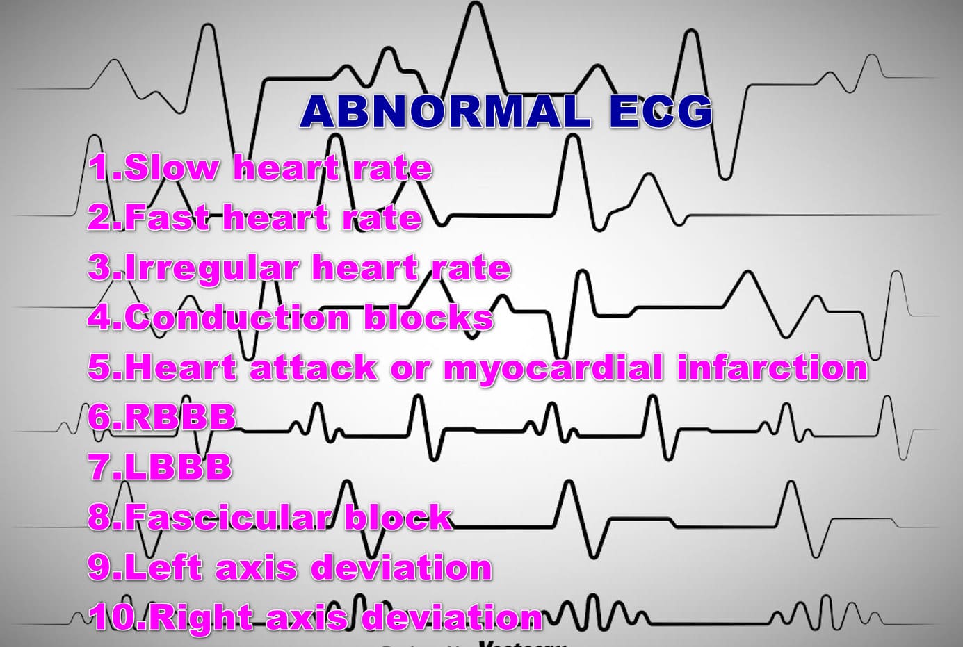
Some ECG abnormalities are
- Slow heart rate
- Fast heart rate
- Irregular heart rate
- Conduction blocks
- Heart attack or myocardial infarction
- RBBB
- LBBB
- Fascicular block
- Left axis deviation
- Right axis deviation








Pingback: Bradycardia or slow heart rate - DM HEART CARE CLINIC